-
Ankle Arthroscopy
Arthroscopy is a surgical procedure during which the internal structure of a joint is examined for diagnosis and treatment of problems inside the joint. Ankle Arthroscopy includes the diagnosis and treatment of ankle conditions. In arthroscopic examination, a small incision is made in the patient's skin through which pencil-sized instruments that have a small lens and lighting system (arthroscope) are passed.
-
Ankle Fracture
Ankle injuries are the most common sports-related injury. An ankle fracture is a break in one or more bones that make up the ankle joint. Sometimes ligaments may also be damaged. Ankle fractures are most often caused by motor vehicle accident, rolling or twisting of ankle, and by tripping or falling.
-
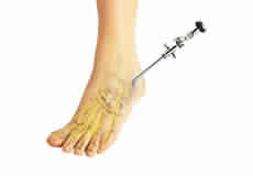
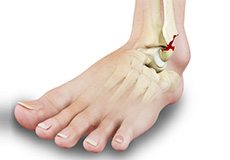
Bunion Surgery
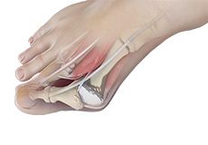
Bunion is a foot deformity that changes the shape of the foot causing the big toe to turn inward, towards the second toe leading to pain and inflammation. A bunion is caused by incorrect footwear, joint damage, arthritis, and genetic disposition.
-
Achilles Tendon Rupture
Achilles tendon is a strong fibrous cord present behind the ankle that connects the calf muscles to heel bone. It is used when you walk, run and jump. When the Achilles tendon becomes thin, weak, or if it is not used, it may be susceptible to injury or damage.
-
Ankle Sprain
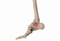
sprain is stretching or tearing of ligaments, which connect adjacent bones in a joint and provides stability to the joint. An ankle sprain is a common injury and occurs when you fall or suddenly twist the ankle joint or when you land your foot in an awkward position after a jump.
-
Common Toe Deformities
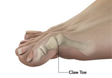
Toes are the digits in your foot and are associated with walking, providing balance, weight-bearing and other activities. A variety of toe deformities occur in children's feet.
-
Achilles Tendon Bursitis
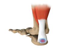
Achilles tendon bursitis or retrocalcaneal bursitis is a condition that commonly occurs in athletes. It is a painful condition caused by swelling of bursa, a fluid-filled sac which is located at the back of the heel under the Achilles tendon.
-
Club Foot
Club foot is a common foot deformity present at birth where in one or both the feet are turned towards an inward and downward position. It is more common in boys than girls. It is also called as talipes equinovarus.
-
High Arch (Cavus Foot)
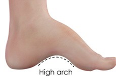
High arch (cavus foot) is a condition in which the arch on the bottom of the foot that runs from the toes to the heel is raised more than normal. Because of this high arch, excessive weight falls on the ball and heel of the foot when walking or standing causing pain and instability.
-
Stress Fracture of the Foot
A stress fracture is described as a small crack in the bone which occurs from an overuse injury of a bone. It commonly develops in the weight bearing bones of the lower leg and foot.
-
Tarsal Coalition
Tarsal coalition is a developmental condition in which there is an abnormal connection between two or more of tarsal bones. Tarsal bones are calcaneus, talus, navicular, and cuboid bones that help in proper functioning of the foot.
-
Tarsal Tunnel Syndrome
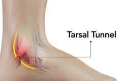
Tarsal tunnel is the gap that is formed between the underlying bones of the foot and the overlying tough fibrous tissue. Tarsal tunnel syndrome refers to a condition where the posterior tibial nerve that lies within the tarsal tunnel is compressed. The condition occurs when the tibial nerve is pinched.
-
Toe Deformities
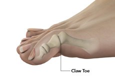
Toes are the digits in your foot and are associated with walking, providing balance, weight-bearing and other activities. A variety of toe deformities occur in children's feet.
What is the Normal Anatomy of the Foot and Ankle?
The foot and ankle form complex joints that are involved in movement and providing stability and balance to the body. The foot and ankle consist of 26 bones, 33 joints, and many muscles, tendons, and ligaments.
Bones of the Ankle
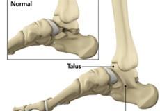
The ankle joint connects the leg with the foot and is composed of three bones: the tibia, fibula, and talus. The tibia or shinbone and fibula or calf bone are bones of the lower leg, which articulate with the talus or ankle bone, enabling up and down movement of the foot.
Three bony bumps present on the ends of the tibia and fibula form parts of the ankle joint:
- The medial malleolus, formed by the tibia, is found on the inside of the ankle.
- The posterior malleolus, also formed by the tibia, is found at the back of the ankle.
- The lateral malleolus, formed by the fibula, is found on the outer aspect of the ankle.
Bones of the Feet
The foot acts as a single functional unit, but can be divided into three parts: the hindfoot, midfoot and forefoot.
The hindfoot forms the ankle and heel, and is made up of the talus bone and calcaneus or heel bone. The heel bone is the largest bone in the foot.
The midfoot connects the hindfoot to the forefoot, and consists of one navicular bone, one cuboid bone, and three cuneiform bones. The navicular bone is found in front of the heel bone, and the cuneiform and cuboid bones are arranged in front of the navicular bone.
These bones are connected to five metatarsal bones of the forefoot that form the arch of the foot for shock absorption while walking or running. The forefoot is also made up of the toes or digits, formed by bones called phalanges - three in each toe, except the big toe, which has only two phalanges. The big toe has two additional tiny round sesamoid bones in the ball of the foot, which helps in upward and downward movements of the toe.
Ankle and Foot Joints
There are 33 joints in the ankle and foot. They include:
- Hinge joints in the ankle, which allow flexion (bending) and extension
- Gliding joints found in the hindfoot, which allow gliding movements
- Condyloid joints found in the forefoot and toes, which allow the flexion (bending) and extension, adduction, and abduction (sideward movement).
The joints of the foot and ankle provide stability and support the weight of your body, helping you to walk or run, and adapt to uneven grounds.
Soft Tissues of the Ankle and Foot
Our feet and ankle bones are held in place and supported by various soft tissues such as cartilage, ligaments, muscles, tendons, and bursae.
The joint surface of all the bones of the ankle and foot are lined by a thin, tough, flexible, and slippery surface called the articular cartilage, which acts as a shock absorber and cushion to reduce friction between the bones. The cartilage is lubricated by synovial fluid, which further enables smooth movement of the bones.
Ligaments are tough rope-like tissue that connect bones to other bones, and hold them in place, providing stability to the joints. The plantar fascia is the largest ligament in the foot, originating from the heel bone to the forefoot, it extends along the lower side of the foot and is involved in maintaining the arch of the foot. The plantar fascia ligament stretches and contracts to provide balance and strength to the foot. Lateral ligaments on the outside of the foot and medial ligaments on the inside of the foot provide stability and allow up and down movement of the foot.
The foot is made up of 20 muscles that help in movement. The main muscles include:
- Anterior tibial muscle, which allows up and down movement of the foot
- Posterior tibial muscle, which supports the arch
- Peroneal tibial muscle, which controls movement on the outside of the ankle
- Extensors, which enable the ankle to raise the toes just before stepping forward
- Flexors, which stabilize the toes against the floor
- Smaller muscles that help the toes to lift and curl
Tendons are soft tissues that connect muscles to bones. The largest and strongest tendon in the foot is the Achilles tendon, present at the back of the lower leg around the heel bone. Other tendons include peroneal and anterior and posterior tibialis.
Bursae are small fluid-filled sacs that decrease friction between tendons and bone or skin. They contain special cells called synovial cells that secrete a lubricating fluid.